
This class runs in two periods and they do not do the same lesson on the same day. You are reading the Period 6A-7B calendar, the one with John Carroll bioethics on Mondays.
Body systems and tissues
Do now
Relate organ systems to tissue types and prepare for the PLTW morgue task.
- Hand in
- Exit ticket: name all four tissue types, one identifying structural feature of each, and one organ that contains each type.
- Where
- Submit this on the class form named on this page. The form requires the student's district Microsoft sign-in. The thank-you page is the submission receipt. A physical handoff counts only when the day page names that route. Doing the activity in myPLTW does not count as submitted.
You get two school days for every day you were absent, so this deadline moves with you.
If the form offers you a saved draft, choose New draft. If the Assignment box comes up empty, type it exactly: Body systems and tissues
If the whole human body is made of only four kinds of , how does a pathologist look at a slice of heart under a microscope and know it failed?
Relate organ systems to types and prepare for the PLTW morgue task.
- • I can name the four types and their roles.
- • I can connect a to the it serves.
- Name any two organs in the human body and guess what they are physically made of.
- A heart pumps and a stomach lining protects. Would you expect those two jobs to be done by the same kind of or different kinds? Why?
- 1Take notes on the four primary types and their functions.
- 2Map two organ systems to the tissues that build them.
- 3Review how a pathologist links damage to .
- 4Complete the PLTW master-the-morgue online task on body systems.
- 5Note the observation variables you will record during microscopy.
What did this day actually feel like?
Body systems and tissues
Four primary tissue types: epithelial, connective, muscle, nervous. Every organ in the body is built from combinations of those four. That is a genuinely surprising fact and it made the body feel much more knowable than it did an hour earlier.
Then how a pathologist reads tissue damage to work out what went wrong. The logic is that you learn what normal looks like so thoroughly that abnormal announces itself.
Turned in: exit ticket → Exit Tickets folder
Fiction. There is no such student. The lessons, labs and dates are the real planned course; the student, the classmates and the conversations are invented.
The same day, drawn.
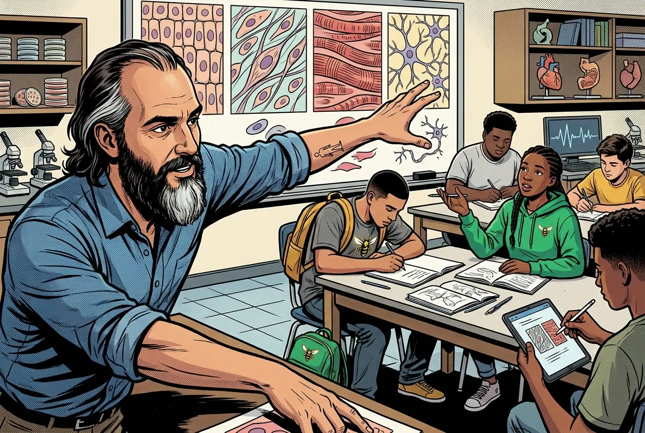
Every organ in the body is built from combinations of just four tissue types. That made the whole body feel more knowable than it had an hour earlier.
Fiction. There is no such student. The lessons, labs and dates are the real planned course; the student, the classmates and the conversations are invented.
🛠 Get unstuck · pick your level
🔑 Today's words · 5
Tap a word in the lesson for a plain meaning and one example. Recycled into next week's Do-Now.
PBS Unit 1 is one case, not twenty topics. A woman was found dead at home, and you are the team that has to work out how she died. You get the evidence in pieces, in the order a real investigation would get it, and you are not told the answer at any point.
Post-mortem interval. You run three independent clocks on a practice case, body cooling, insect accumulated degree hours, and rigor with lividity, then report the overlap between them instead of one time.
- Activity 1.2.1 Anatomy of an Autopsy
- Activity 1.2.2 Time of Death
Look this up in myPLTW to check you are in the right place.
What you cannot claim in this case, on any day
- A physiological chart records breathing and heart rate. It does not measure lying, and nothing in this unit produces a deception call.
- Microscopic hair comparison can support consistency or support exclusion. It cannot identify a person.
- A fingerprint pattern class can narrow a field or exclude a person. By itself it cannot name a source, and it never says when or why a finger touched something.
- A scheduled event is not proof that a person was there. Time of death is not established by the early evidence.
- Repeating a measurement reduces random error. It does not remove a calibration offset, a shared bias, contamination, or a weak method.
Where the case record itself lives: the case file, the source packet and the timeline are PLTW materials and are handed out on paper in class. They are not posted here.
Do the work · 80-minute blockfirst 5 min = hook▸
💡 Big idea: A pathologist studies just four types instead of dozens of organs because every organ is built from those four, so learning how each tissue breaks lets you read damage anywhere in the body.
- 0:00Quick-draw warm-up: sketch what you think a cell looks like; compare to actual cell diagrams
- 0:08Teacher-led notes: four types, defining features of each, which organs they build
- 0:28Mapping activity: draw two organ systems and label which types make up each structure
- 0:42myPLTW: complete the master-the-morgue body-systems online task
- 1:02Write observation variables for Wednesday's microscopy: what will you record for each slide?
- 1:10Exit ticket: name the four types and one identifying feature of each
- • Everything in your body, every organ, every gland, every vessel, is made from combinations of just four types. If you can identify the tissue, you can start to understand what organ it came from and what might have gone wrong with it.
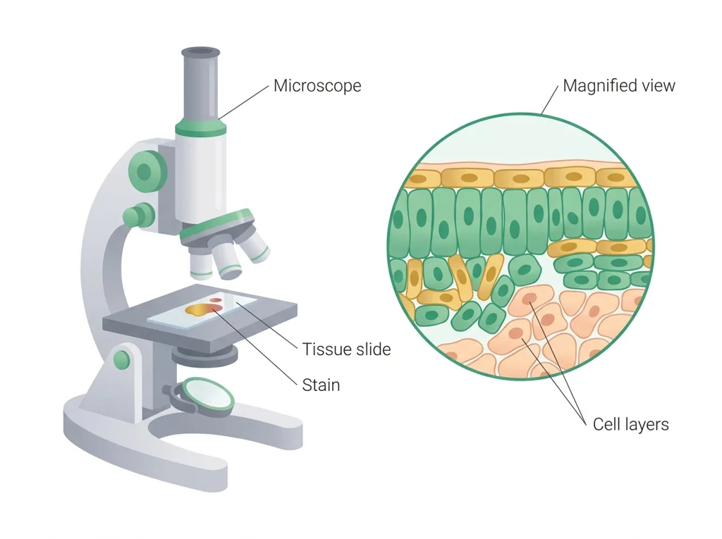
- • Pathologists use , the microscopic study of , to determine . They are looking for patterns: cells that are swollen, dead, or abnormally shaped. Those patterns are the clues.
- • Tomorrow you will identify types on real slides and examine a preserved heart. Today we build the knowledge you need to actually recognize what you are looking at.
- • By the end of today you should be able to look at a cross-section of and at least ask the right question: , , muscle, or nerve?
- • The four primary types are (covering/lining), (support/binding), muscle (movement), and nervous (communication).
- • A pathologist identifies by looking for -level changes such as necrosis, inflammation, or structural damage in slides.
- • Organ systems are not independent: the cardiovascular system depends on muscular and tissue, and a failure in one tissue type can cascade across systems.
Unit 1.2 Master the Morgue: Body systems, toxicology evidence, tissue microscopy, gross anatomy, preserved-heart/autopsy alternative. · Body systems and tissues
Day 1 of this lesson. Open this exact section in myPLTW (find it in Clever, Microsoft sign-in), then do the work below.
Do this: In myPLTW, open Lesson 1.2 Master the Morgue and go to Activity 1.2.1 of an and Activity 1.2.2 Time of Death. Finish the body-systems questions in the first one, then start the time-of-death work in the second.
Mark Activity 1.2.1 of an and Activity 1.2.2 Time of Death complete in myPLTW.
You read the Lesson 1.2 overview Monday. By the end of today the myPLTW body-systems task should be finished.
Screenshot of myPLTW showing Activity 1.2.1 of an and Activity 1.2.2 Time of Death marked complete.
The official PLTW activity stays inside myPLTW. If myPLTW will not open, use F1 and E1-E3 on this page to complete today's local evidence decision, then make up the official activity when access returns. Submit this on the class form named on this page. The form requires the student's district Microsoft sign-in. The thank-you page is the submission receipt. A physical handoff counts only when the day page names that route. Doing the activity in myPLTW does not count as submitted.
Check things off as you work, then submit. This tells Mr. Mendoza how you're doing so he can help the class. It does not replace turning in your producible through the submission route shown below.
Use the code Mr. Mendoza gave you, not your name. Saved on this device.
Unit 1.2 Master the Morgue: Body systems, toxicology evidence, tissue microscopy, gross anatomy, preserved-heart/autopsy alternative. · Body systems and tissues
In myPLTW, open Lesson 1.2 Master the Morgue and go to Activity 1.2.1 of an and Activity 1.2.2 Time of Death. Finish the body-systems questions in the first one, then start the time-of-death work in the second.
You read the Lesson 1.2 overview Monday. By the end of today the myPLTW body-systems task should be finished.
This is how Mr. Mendoza sees the class keeping pace with PLTW. Be honest, it only helps if it is accurate.
🎯 Relate organ systems to types and prepare for the PLTW morgue task.
- Take notes on the four primary types and their functions.
- Map two organ systems to the tissues that build them.
- Review how a pathologist links damage to .
- Complete the PLTW master-the-morgue online task on body systems.
- Note the observation variables you will record during microscopy.
Exit ticket: Exit ticket: name all four types, one identifying structural feature of each, and one organ that contains each type.
Submit this on the class form named on this page. The form requires the student's district Microsoft sign-in. The thank-you page is the submission receipt. A physical handoff counts only when the day page names that route. Doing the activity in myPLTW does not count as submitted. Use the checklist just below and upload by 11:29 PM for full credit. Absent with an excused absence? You get two school days for every day you were absent, so this deadline moves with you.
| Task | Who |
|---|---|
| Take notes on the four primary types and their functions. | _______ |
| Map two organ systems to the tissues that build them. | _______ |
| Review how a pathologist links damage to . | _______ |
| Complete the PLTW master-the-morgue online task on body systems. | _______ |
| Note the observation variables you will record during microscopy. | _______ |
Working solo? Put your own name in "Who" for every row.
- I can name the four types and their roles.
- I can connect a to the it serves.
- 1Do thisRelate organ systems to tissue types and prepare for the PLTW morgue task.
- 2Use this resource
- 3Submit thisExit ticket: Exit ticket: name all four tissue types, one identifying structural feature of each, and one organ that contains each type.
- 4Submit it here
- 1Open the form. It must say For students at the top: if it says For parents and guardians, press Back and pick I am the student.
- 2If it offers you a saved draft, choose New draft: an old draft brings back the old assignment.
- 3Sign in with your district Microsoft account, not a personal one.
- 4Check the Assignment box says today's assignment from this page, then pick your period and type your student ID, all nine digits.
- 5Attach your file and press Submit. The thank-you page is your receipt.
Submit this on the class form named on this page. The form requires the student's district Microsoft sign-in. The thank-you page is the submission receipt. A physical handoff counts only when the day page names that route. Doing the activity in myPLTW does not count as submitted. Principles of Biomedical Technology (Principles of Biomedical Science) › Unit 1.2 Master the Morgue: Body systems, toxicology evidence, tissue microscopy, gross anatomy, preserved-heart/autopsy alternative. › Exit ticketTurn it in
Learn it · deck, reading, and vocabulary▸
The deck carries the prior idea forward, lets you inspect an analogy, maps the rule to biology, and ends with the same evidence decision and exit ticket used on this page.
Generated from this lesson's canonical data with a red-team citation check.
Should testing for Novel Flu Syndrome be mandatory?
A pathologist studies just four types instead of dozens of organs because every organ is built from those four, so learning how each tissue breaks lets you read damage anywhere in the body.
A mechanic studies a tool whose shape allows one job but limits another.
- Which feature makes the tool work?
- What changes if that feature bends or breaks?
- Which observation shows function rather than appearance?
Structure creates possibilities and limits for function.
Living tissues adapt and interact with other systems; a metal tool does not.
- • Tool shape maps to .
- • The job maps to physiological function.
- • Damage maps to a predicted functional change.
Driving question: If the whole human body is made of only four kinds of , how does a pathologist look at a slice of heart under a microscope and know it failed?
What you already know: Should testing for Novel Flu Syndrome be mandatory?
New idea: A pathologist studies just four types instead of dozens of organs because every organ is built from those four, so learning how each tissue breaks lets you read damage anywhere in the body.
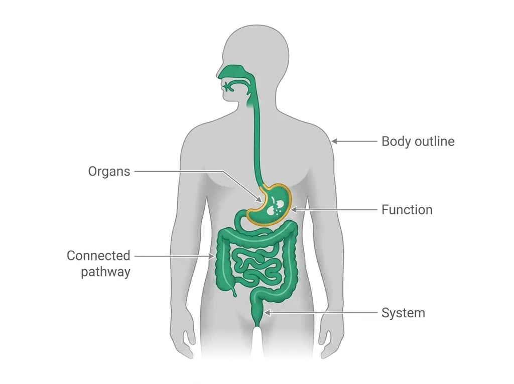
Visual or model: F1. F1. A lesson illustration or teaching diagram for Body systems and tissues. Use it with E1-E3; it is a model or context image, not experimental or patient data. What to notice: Use the labels and arrows in F1 to identify the relationship that supports Body systems and tissues.
- Observe or measure the relevant feature in today's lesson.
- Organize the observation with a stable evidence ID.
- Apply this rule: Structure creates possibilities and limits for function.
- Choose the option the evidence supports and state the limit of the conclusion.
Real biomedical example: If the whole human body is made of only four kinds of , how does a pathologist look at a slice of heart under a microscope and know it failed?
What the evidence supports: E1-E3 and F1 support the daily take-home when the response meets the stated success criteria.
What it cannot prove: The package does not support claims beyond this lesson's or any real patient diagnosis.
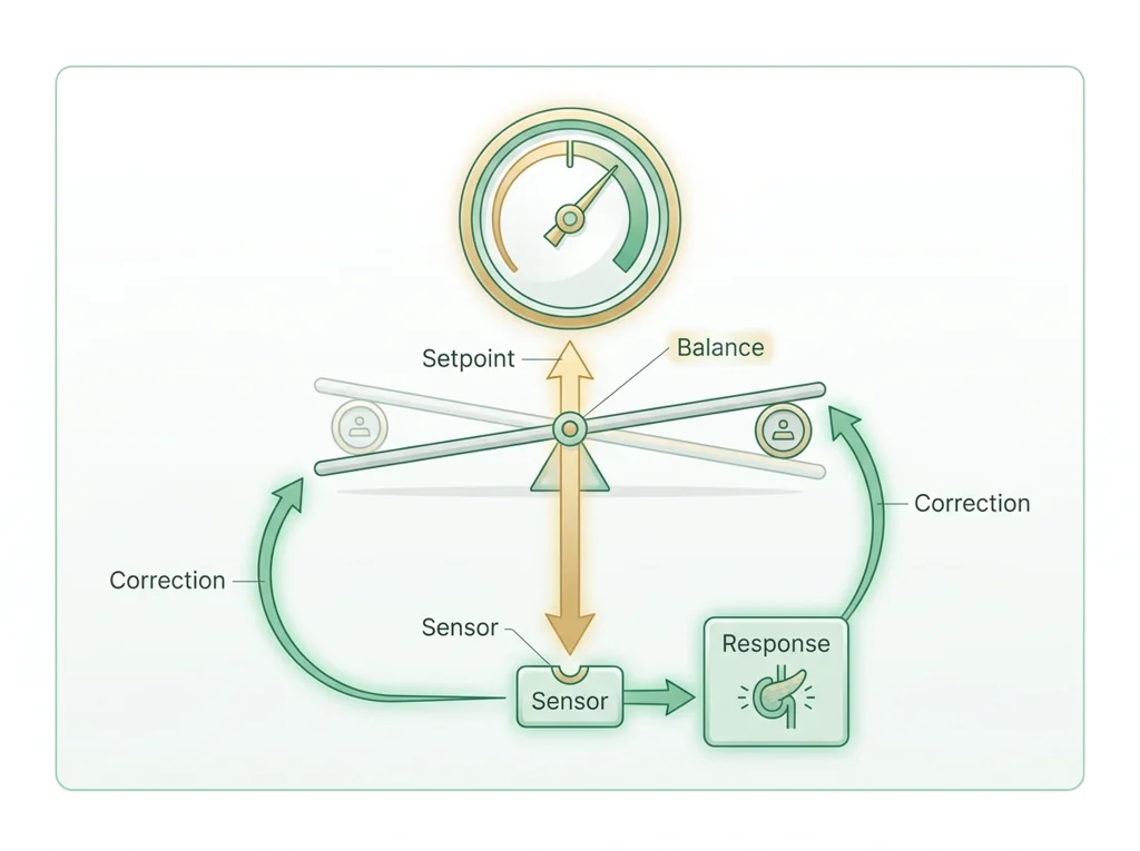
- • : The body's ongoing process of keeping internal conditions like temperature, blood sugar, and pH steady despite changes in the outside environment.
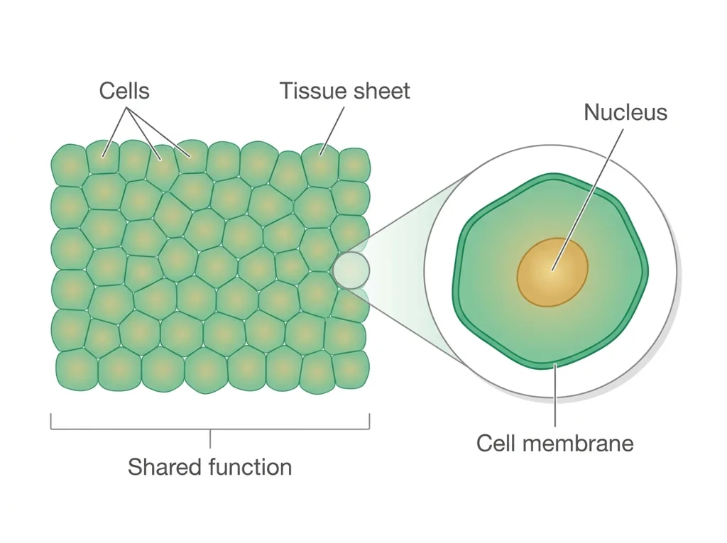
- • : A group of similar cells working together to perform a shared function, such as muscle, nerve, or .
- • : A group of organs that work together to carry out a major body function, such as the digestive or circulatory system.
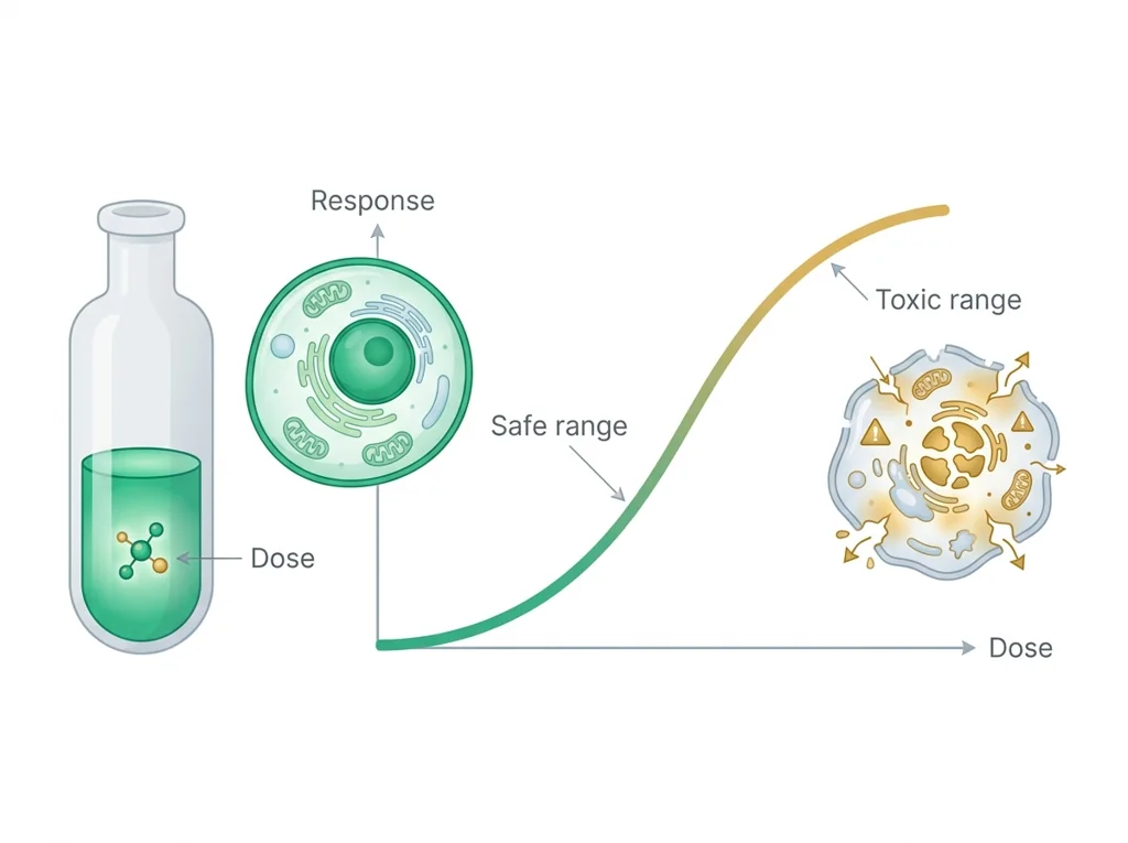
- • toxicology: The science of how chemicals and other substances cause harm to living things, including the dose at which they become dangerous.
- • : The study of the microscopic structure of tissues, examining thin stained slices under a microscope to see how cells are arranged.
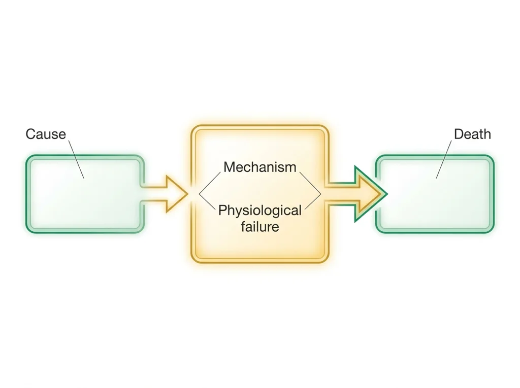
- • : The specific physiological breakdown inside the body that directly ends life, such as a fatal heart rhythm or blood loss, separate from the underlying cause.
Use it now: Choose one decision option. Cite E1 and E3, then explain how the rule connects the evidence to your choice.
Go further, optional: The source links below are optional enrichment. Every fact required for today's local evidence decision appears in this lesson package.
Standard anatomical terms describe location and structure precisely, while physiology describes function; connecting levels from cells to tissues, organs, and systems supports a bounded explanation.
Limit: A classroom map or organization model simplifies three-dimensional variation and cannot diagnose injury or disease.
Structure creates possibilities and limits for function.
Limit: Living tissues adapt and interact with other systems; a metal tool does not.
You can name the four types and their roles.
Limit: E3 defines the classroom product or success criterion. It is not independent scientific evidence and cannot justify a clinical or causal claim.
PLTW-PBT@P67-2026-09-23 · Simulated classroom evidence scenario
Your role: biomedical team member
Decision: Your team must decide what the evidence from today's lesson supports before submitting the exit response named on the lesson page.
- • Select the option best supported by E1-E3.
- • Select a reasonable alternative and name the evidence it would require.
- • Delay the claim because the evidence does not distinguish the options.
Response: State one choice, cite at least two evidence IDs, explain the rule that connects them, and add one limitation. Submit it as the exit response.
Claim ceiling: Today's evidence supports a classroom claim about today's lesson. It cannot prove causation, diagnose a real patient, or justify action outside this room.
Reason for review: Your team must decide what the evidence from today's lesson supports before submitting the exit response named on the lesson page.
Context: Every organ is built from just four types, so damage at the tissue level is the evidence a pathologist uses to explain a death.
- • T1: Take notes on the four primary types and their functions.
- • T2: Map two organ systems to the tissues that build them.
- • T3: Review how a pathologist links damage to .
- • T4: Complete the PLTW master-the-morgue online task on body systems.
- • T5: Note the observation variables you will record during microscopy.
- • E1: Standard anatomical terms describe location and structure precisely, while physiology describes function; connecting levels from cells to tissues, organs, and systems supports a bounded explanation.
- • E2: Structure creates possibilities and limits for function.
- • E3: You can name the four types and their roles.
Measurements: No patient measurement is supplied unless it appears explicitly in E1-E3 or F1. Do not invent a value.
Figure finding: Teaching diagram for Body systems and tissues. Use the labels and arrows to identify the decision-relevant relationship. This is a teaching model, not patient or experimental data.
Uncertainty: This is a composite classroom scenario. Missing history, measurements, or confirmation tests remain unknown and limit the conclusion.
Students often think Students think each organ is made of its own unique special material, so the heart is made of heart stuff and the liver is made of liver stuff.. The trap: That is a trap because all organs are combinations of the same four types (, , muscle, nervous); the heart and the liver share tissues, which is why a single tissue failure can cascade across many different organs.
Four tissue types, a feature, and an organ:
- Epithelial: tightly packed cells that cover and line surfaces; found in the skin.
- Connective: cells spread in a matrix that supports and binds; found in bone.
- Muscle: long fibers that contract; found in the heart.
- Nervous: branching cells that send signals; found in the brain.
Why it matters: organs are built from combinations of these four, so recognizing them is the first step a pathologist takes when reading a slide.
| Tissue type | Feature | Organ example |
|---|---|---|
| Epithelial | Covers and lines surfaces | Skin |
| Connective | Matrix supports and binds | Bone |
| Muscle | Fibers that contract | Heart |
| Nervous | Cells that send signals | Brain |
This model shows the level of evidence and organization needed to complete: An exit ticket naming the four primary tissue types, one structural feature of each, and one organ that contains each, checking recall before microscopy.
- Name the prompt or task.
- Answer it directly with the key evidence.
- Check that the response matches the requested format.
Keep the structure. Replace the question, facts, measurements, and evidence. Then recheck units, vocabulary, and whether the conclusion goes beyond the evidence.
Also due today: Hand in your exit ticket before leaving class. Also complete the online master-the-morgue body-systems task.
- CER:
- Claim, Evidence, Reasoning: make a claim, back it with evidence, explain your reasoning.
- SOP:
- Standard Operating Procedure, the exact steps to follow (especially in a lab).
- Tracker:
- Your PLTW progress log where you record completed evidence.
- myPLTW:
- The PLTW course site where you do the online activities. Find it in Clever with your Microsoft sign-in, right next to Schoology.
Tap the speaker to hear a term. Add two of these to your notebook glossary with a definition and an example in your own words.
Pick just 2 or 3 words from today and make them yours: write what each one means in your own words, name the context clue or evidence that helped, then give one example from what you actually did in Body systems and tissues. Try your own words first; the glossary is there if you get stuck. This is voluntary and counts as extra credit, so keep it short.
Saved on this device. Show Mr. Mendoza or add these to your notebook glossary to claim the extra credit.
Hand-picked readings and interactives for this lesson, from authoritative open organizations and PLTW's own public course outline.
Check yourself · commit, then reveal▸
Claim ceiling for this check: Today's evidence supports a classroom claim about today's lesson. It cannot prove causation, diagnose a real patient, or justify action outside this room.
The wall of the heart is mostly one tissue type doing one job. Which of the four tissue types is it, and what job is that tissue built to do?
Write an answer and pick a confidence to unlock the key.
Fast retrieval with instant answers, not the commit-then-reveal check above. Try each from memory first: write what you remember about the earlier units, then check yourself here.
Go further and get help▸
Run this before you touch the bench. It is built from the real lab procedure, so the decisions you make here are the ones you will make with the equipment in your hands. This lesson has more than one, and they cover different skills.
What today's skills lead to. These are real health-science careers this course builds toward. Tap one to see, on the US Department of Labor's O*NET site, what the job actually involves, what it pays, and how fast it is growing.
The bench work needs equipment you do not have at home. Do the thinking half now: read the procedure, write your prediction, and set up your data table so it is ready.
Back in class. Ask Mr. Mendoza for the class data set, or for a bench slot to run it yourself. Do not submit a Exit ticket with invented numbers.
Class still runs. Complete the online activity above (it's self-guided). Need the concept taught without a teacher? Use this authoritative explainer:
Khan Academy: human body systems (Health and medicine)- CompleteEvery required part of the artifact is present, nothing left blank.
- AccurateThe science and the data are correct and match the evidence.
- Scientific reasoningYou explain your claim with evidence and reasoning (CER), not just an answer.
- Professional communicationClear, organized, labeled, and written the way a clinician or scientist would.
- SubmittedTurned in through the one route named under Submit here and confirmed by the form receipt or the named physical handoff. Not in Schoology: that is where the report-card grade appears later.