
Brain dissection or virtual
Safety gate · before any work
- This is a sheep-brain dissection. If you are working with a real preserved specimen, wear gloves, goggles, and an apron for the entire lab, and keep them on until the specimen is back in the tray, your station is cleaned, and your hands are washed.
- Preserved specimens are stored in a fixative (often formaldehyde or formalin, sometimes a phenoxyethanol-based solution) that gives off fumes which sting the eyes, nose, and throat and can irritate the skin. Work in a ventilated space, keep the specimen in its tray, do not lean in close to breathe over it, and rinse any preserving fluid off your gloves in the sink your teacher points you to. Tell your teacher right away if fumes make you dizzy, your eyes water, or fluid gets on bare skin or in your eyes, and flush skin or eyes with water for several minutes if that happens.
- Scalpels and probes are sharp. Use them only when your teacher directs, cut and probe away from your own hand and from your lab partner, make one slow coronal (front-to-back) cut for the internal view, and never pass an open blade hand to hand. Tell your teacher about any cut right away so it can be washed and covered.
Do now
Examine brain regions through a sheep-brain dissection or a virtual brain.
- Hand in
- Labeled brain-region map identifying cerebrum, cerebellum, and brainstem with one function each, plus gray and white matter boundary marked on an internal view.
- Where
- Turn this in at the drop folder with your district Microsoft sign-in, or hand it to Mr. Mendoza in class. Both count as submitted. Doing the activity in myPLTW does not.
You get two school days for every day you were absent, so this deadline moves with you.
Holding a real sheep brain, can you find the region that controls balance, and predict what would go wrong if a patient injured that exact spot?
Examine brain regions through a sheep-brain dissection or a virtual brain.
- • You can locate the , cerebellum, and brainstem.
- • You can state one function for each region.
- Name one thing your brain controls right now without you thinking about it.
- If someone's balance is wrecked but their thinking is sharp, which brain region would you check first?
- 1Review the and handling steps for the sheep-brain or virtual model.
- 2Identify the , cerebellum, and brainstem on the .
- 3Note one function controlled by each region you identify.
- 4Compare the external and a cut internal view to locate gray and white matter.
- 5Submit your labeled brain-region map with functions.
What did this day actually feel like?
Brain dissection
DISSECTION Sheep brain. I was not sure how I would handle this and I was fine, which surprised me. Cerebrum, cerebellum, brainstem, and the texture difference between grey and white matter, which is obvious in your hands and invisible in a textbook.
One person in our group could not do it and Mr. Mendoza gave them the virtual version without making it a thing. I noticed that.
Turned in: labeled brain region map → Lab Reports folder
Fiction. There is no such student. The lessons, labs and dates are the real planned course; the student, the classmates and the conversations are invented.
The same day, drawn.
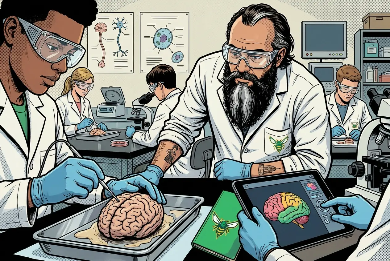
Brain day. The virtual option is right there and nobody has to explain why they took it. I took the specimen and my hands were not steady.
MR. MENDOZA
Take the virtual version. Nobody has to explain themselves for that.
Fiction. There is no such student. The lessons, labs and dates are the real planned course; the student, the classmates and the conversations are invented.
🛠 Get unstuck · pick your level
Lab day: Tier 1 is the whole class at the bench. No extension today.
🔑 Today's words · 5
Tap a word in the lesson for a plain meaning and one example. Recycled into next week's Do-Now.
Do the work · 80-minute blockfirst 5 min = hook▸
💡 Big idea: Each brain region runs a different set of functions, so where an injury lands determines which specific ability a patient loses.
- 0-10 briefing and handling protocol for sheep brain or virtual login
- 10-25External examination: identify , cerebellum, brainstem; note one function each
- 25-45Internal (coronal) cut: locate gray matter and white matter
- 45-60Draw and label brain-region map: three regions with functions; gray/white matter boundary
- 60-75Group comparison: does your map match the reference? correct discrepancies
- 75-80Submit labeled map; clean up or log off virtual
- • Today you examine a real or virtual brain. This is the most direct way to understand why region location matters.
- • You are looking for three regions and the boundary between gray and white matter. Those four observations go on your brain-region map.
- • Handle the with care and with dignity. This is a real nervous system that ran a real animal for years.
- • Your map today becomes the reference for Thursday when we connect those regions to the CNS and PNS organization.
- • : largest region; controls voluntary movement, sensory processing, language, memory, and higher cognition. Divided into four lobes (, parietal, temporal, occipital).
- • Cerebellum: located to the brainstem; coordinates balance, fine motor control, and learned movement sequences.
- • Brainstem (medulla oblongata, pons, midbrain): controls autonomic functions including heart rate, , and ; connects brain to spinal cord.
- • Gray matter contains cell bodies; white matter contains myelinated axons. On a cut surface, gray is darker and peripheral, white is lighter and central (in the ).
Unit 2.1 Getting Nervous: Nervous system structure, brain anatomy, neurons, signaling, sheep brain or virtual alternative. · Brain dissection or virtual
Day 3 of this lesson. Open this exact section in myPLTW (find it in Clever, Microsoft sign-in), then do the work below.
Do this: Complete the brain-regions task in Lesson 2.1 Getting Nervous on myPLTW that accompanies today's dissection or virtual brain; match structures to functions.
Mark the brain-regions task complete after submitting your dissection notes or virtual-brain annotation.
task is done; today the brain-regions task should show complete.
myPLTW completion status plus submitted annotation.
The official PLTW activity stays inside myPLTW. If myPLTW will not open, use F1 and E1-E3 on this page to complete today's local evidence decision, then make up the official activity when access returns. Turn this in at the drop folder with your district Microsoft sign-in, or hand it to Mr. Mendoza in class. Both count as submitted. Doing the activity in myPLTW does not.
Check things off as you work, then submit. This tells Mr. Mendoza how you're doing so he can help the class. It does not replace turning in your producible through the submission route shown below.
Use the code Mr. Mendoza gave you, not your name. Saved on this device.
Unit 2.1 Getting Nervous: Nervous system structure, brain anatomy, neurons, signaling, sheep brain or virtual alternative. · Brain dissection or virtual
Complete the brain-regions task in Lesson 2.1 Getting Nervous on myPLTW that accompanies today's dissection or virtual brain; match structures to functions.
task is done; today the brain-regions task should show complete.
This is how Mr. Mendoza sees the class keeping pace with PLTW. Be honest, it only helps if it is accurate.
🎯 Examine brain regions through a sheep-brain dissection or a virtual brain.
- Review the and handling steps for the sheep-brain or virtual model.
- Identify the , cerebellum, and brainstem on the .
- Note one function controlled by each region you identify.
- Compare the external and a cut internal view to locate gray and white matter.
- Submit your labeled brain-region map with functions.
Lab report: Labeled brain-region map identifying , cerebellum, and brainstem with one function each, plus gray and white matter boundary marked on an internal view.
Turn this in at the drop folder with your district Microsoft sign-in, or hand it to Mr. Mendoza in class. Both count as submitted. Doing the activity in myPLTW does not. Use the checklist just below and upload by 11:29 PM for full credit. Absent with an excused absence? You get two school days for every day you were absent, so this deadline moves with you.
| Task | Who |
|---|---|
| Review the and handling steps for the sheep-brain or virtual model. | _______ |
| Identify the , cerebellum, and brainstem on the . | _______ |
| Note one function controlled by each region you identify. | _______ |
| Compare the external and a cut internal view to locate gray and white matter. | _______ |
| Submit your labeled brain-region map with functions. | _______ |
Working solo? Put your own name in "Who" for every row.
- You can locate the , cerebellum, and brainstem.
- You can state one function for each region.
- 1Do thisExamine brain regions through a sheep-brain dissection or a virtual brain.
- 2Use this resource
- 3Submit thisLab report: Labeled brain-region map identifying cerebrum, cerebellum, and brainstem with one function each, plus gray and white matter boundary marked on an internal view.
- 4Submit it here
- 1Open the drop folder.
- 2Sign in with your district Microsoft account, not a personal one.
- 3Upload the file, named Lastname_Firstname__Assignment Title.
- 4Your own upload panel says Uploaded with a green check: that is your receipt.
Turn this in at the drop folder with your district Microsoft sign-in, or hand it to Mr. Mendoza in class. Both count as submitted. Doing the activity in myPLTW does not. Human Anatomy & Physiology (Human Body Systems) › Unit 2.1 Getting Nervous: Nervous system structure, brain anatomy, neurons, signaling, sheep brain or virtual alternative. › Lab reportOpen the drop folder
Learn it · deck, reading, and vocabulary▸
The deck carries the prior idea forward, lets you inspect an analogy, maps the rule to biology, and ends with the same evidence decision and exit ticket used on this page.
Generated from this lesson's canonical data with a red-team citation check.
Neuroimaging shows brain structure and activity but not future choices, so using scan data to predict behavior without consent trades away individual autonomy for a promise the technology cannot keep.
Each brain region runs a different set of functions, so where an injury lands determines which specific ability a patient loses.
A mechanic studies a tool whose shape allows one job but limits another.
- Which feature makes the tool work?
- What changes if that feature bends or breaks?
- Which observation shows function rather than appearance?
Structure creates possibilities and limits for function.
Living tissues adapt and interact with other systems; a metal tool does not.
- • Tool shape maps to .
- • The job maps to physiological function.
- • Damage maps to a predicted functional change.
Driving question: Holding a real sheep brain, can you find the region that controls balance, and predict what would go wrong if a patient injured that exact spot?
What you already know: Neuroimaging shows brain structure and activity but not future choices, so using scan data to predict behavior without consent trades away individual autonomy for a promise the technology cannot keep.
New idea: Each brain region runs a different set of functions, so where an injury lands determines which specific ability a patient loses.
Visual or model: F1. F1. A lesson illustration or teaching diagram for Brain dissection or virtual. Use it with E1-E3; it is a model or context image, not experimental or patient data. What to notice: Trace the labeled structure, movement, or system relationship that connects form to function.
- Observe or measure the relevant feature in Brain dissection or virtual.
- Organize the observation with a stable evidence ID.
- Apply this rule: Structure creates possibilities and limits for function.
- Choose the option the evidence supports and state the limit of the conclusion.
Real biomedical example: Holding a real sheep brain, can you find the region that controls balance, and predict what would go wrong if a patient injured that exact spot?
What the evidence supports: E1-E3 and F1 support the daily take-home when the response meets the stated success criteria.
What it cannot prove: The package does not support claims beyond this lesson's or any real patient diagnosis.
- • : A nerve cell that carries electrical and chemical signals, with branches that receive input and a long fiber that sends the signal onward.
- • : A branching extension of a that receives incoming signals from other nerve cells and carries them toward the cell body.
- • axon: The long, slender fiber of a that carries electrical signals away from the cell body toward other cells.
- • : The tiny junction where one passes a signal to the next, usually by releasing chemical messengers across a small gap.
- • : A chemical messenger released by a that crosses the to pass a signal to the next neuron, muscle, or gland.
- • CNS: The central nervous system, made of the brain and spinal cord, which processes information and directs the body's responses.
- • PNS: The peripheral nervous system: all the nerves outside the brain and spinal cord that carry signals between the body and the central nervous system.
- • : The largest part of the brain, split into two hemispheres, that handles thinking, voluntary movement, sensation, language, and memory.
Use it now: Choose one decision option. Cite E1 and E3, then explain how the rule connects the evidence to your choice.
Go further, optional: The source links below are optional enrichment. Every fact required for today's local evidence decision appears in this lesson package.
: largest region; controls voluntary movement, sensory processing, language, memory, and higher cognition. Divided into four lobes (, parietal, temporal, occipital).
Limit: E1 supplies context or an observation; it does not by itself establish the explanation.
Each brain region runs a different set of functions, so where an injury lands determines which specific ability a patient loses.
Limit: E2 is a teaching statement or comparison and must be checked against the task evidence.
You can locate the , cerebellum, and brainstem.
Limit: E3 supports only the result or product criterion named here; it cannot justify a broader clinical or causal claim.
PLTW-HAP-2027-03-11 · Simulated classroom evidence scenario
Your role: anatomy and physiology consultant
Decision: Your team must decide what the evidence from Brain dissection or virtual supports before submitting the lab report named on the lesson page.
- • Proceed because the readiness evidence is complete.
- • Pause and correct the named setup or gap.
- • Repeat the measurement because quality controls are not acceptable.
Response: State one choice, cite at least two evidence IDs, explain the rule that connects them, and add one limitation. Submit it as the lab report.
Claim ceiling: The supplied lesson evidence can support an observation, pattern, classroom mechanism, or next-step decision about Brain dissection or virtual. It cannot by itself prove causation, establish a real clinical diagnosis, or justify action outside this classroom task.
Reason for review: Your team must decide what the evidence from Brain dissection or virtual supports before submitting the lab report named on the lesson page.
Context: The brain divides labor by region, so where an injury happens predicts which function is lost, which is why is the first clue in every neurological case.
- • T1: Review the and handling steps for the sheep-brain or virtual model.
- • T2: Identify the , cerebellum, and brainstem on the .
- • T3: Note one function controlled by each region you identify.
- • T4: Compare the external and a cut internal view to locate gray and white matter.
- • T5: Submit your labeled brain-region map with functions.
- • E1: : largest region; controls voluntary movement, sensory processing, language, memory, and higher cognition. Divided into four lobes (, parietal, temporal, occipital).
- • E2: Each brain region runs a different set of functions, so where an injury lands determines which specific ability a patient loses.
- • E3: You can locate the , cerebellum, and brainstem.
Measurements: Use only the measurements, units, graph, or counts supplied in today's task. No additional patient measurement is implied.
Figure finding: Teaching diagram for Brain dissection or virtual. Trace the labeled structure, movement, or system relationship that connects form to function. This is a teaching model, not patient or experimental data.
Uncertainty: This is a composite classroom scenario. Missing history, measurements, or confirmation tests remain unknown and limit the conclusion.
Rate or percent = part / comparison total x 100%. Percent change = (new - comparison) / comparison x 100%.
If 18 of 60 records meet a condition, the frequency is 18 / 60 x 100% = 30%.
Name the comparison total. A percent describes the supplied group and does not automatically predict an individual's outcome.
Use today's supplied counts to calculate one rate, risk, frequency, or percent change. Show the denominator and interpretation.
- • The solution must address the stated need in Brain dissection or virtual.
- • The decision must be supported by E1-E3.
- • The final product must make the success criteria visible.
- • Complete the work inside the 80-minute block.
- • Use only supplied or teacher-approved materials and evidence.
- • Do not trade , accessibility, or privacy for speed.
- • and evidence quality: must pass before scoring other criteria.
- • User need and effectiveness: highest scored criterion.
- • Time, cost, and ease of use: compare only after and effectiveness pass.
Test evidence: For each option, record the E1-E3 result that supports or fails each criterion. Do not assign a score without a named observation.
- Version or option tested
- Criterion met or missed
- Evidence ID and result
- Revision made
- Reason for the revision
- Need and user
- Criteria and constraints
- Chosen option and evidence
- Test result
- Revision and reason
Students often think Students often think the whole brain works on every task together, so any injury just makes you 'a little worse at everything.'. The trap: That is the trap. Regions specialize: the cerebellum handles balance, the brainstem runs heart rate and breathing, the handles thought and voluntary movement. That is why a specific injury causes a specific deficit, not a general dimming.
Brain-region map (external and one cut view):
- Cerebrum: largest region, top and front. Function: voluntary movement and higher thinking (one example: language).
- Cerebellum: smaller, wrinkled region at the lower back. Function: balance and fine motor coordination.
- Brainstem: stalk connecting to the spinal cord. Function: automatic survival controls (one example: breathing rate).
Internal (cut) view note: In the cerebrum, gray matter is the darker outer layer (neuron cell bodies) and white matter is the lighter inner region (myelinated axons). I marked the boundary where the darker outer band meets the lighter core.
| Region | Location | One function |
|---|---|---|
| Cerebrum | Top and front, largest | Voluntary movement and language |
| Cerebellum | Lower back, wrinkled | Balance and fine motor coordination |
| Brainstem | Stalk to spinal cord | Breathing and heart rate |
This model shows the level of evidence and organization needed to complete: A labeled map of the brain (from a sheep-brain dissection or a virtual model) identifying the cerebrum, cerebellum, and brainstem with one function each, and marking the gray and white matter boundary on a cut internal view.
- State the question and method.
- Present the observations and data with units.
- Explain the result, limitations, and next investigation.
Keep the structure. Replace the question, facts, measurements, and evidence. Then recheck units, vocabulary, and whether the conclusion goes beyond the evidence.
Also due today: Submit your completed brain-region map.
- CER:
- Claim, Evidence, Reasoning: make a claim, back it with evidence, explain your reasoning.
- SOP:
- Standard Operating Procedure, the exact steps to follow (especially in a lab).
- Tracker:
- Your PLTW progress log where you record completed evidence.
- myPLTW:
- The PLTW course site where you do the online activities. Find it in Clever with your Microsoft sign-in, right next to Schoology.
Tap the speaker to hear a term. Add two of these to your notebook glossary with a definition and an example in your own words.
Pick just 2 or 3 words from today and make them yours: write what each one means in your own words, name the context clue or evidence that helped, then give one example from what you actually did in Brain dissection or virtual. Try your own words first; the glossary is there if you get stuck. This is voluntary and counts as extra credit, so keep it short.
Saved on this device. Show Mr. Mendoza or add these to your notebook glossary to claim the extra credit.
Hand-picked readings, videos, and interactives for this lesson, all free and from authoritative open organizations (NIH, CDC, OpenStax, Khan Academy, PhET, HHMI, and more).
A fillable, Cornell-style notebook for Unit 2: Research Ready. Type your notes, cues, and summaries right in the PDF, or print it and write by hand. Each lesson page has a cue column, a notes column, and a summary box, plus dated lab-record pages you can turn in.
HBS Unit 2 notebook: Research Ready Fillable PDFCornell notes + lab recordsOpenVetted readings and references for this unit. Use them to prepare, to catch up if you were absent, or to go deeper on today's target.
This site may ask you to verify you are human. If it blocks you on a school device, ask for the class copy.
Check yourself · commit, then reveal▸
Claim ceiling for this check: The supplied lesson evidence can support an observation, pattern, classroom mechanism, or next-step decision about Brain dissection or virtual. It cannot by itself prove causation, establish a real clinical diagnosis, or justify action outside this classroom task.
On a cut surface of the cerebrum, is the darker gray matter on the outside or the inside, and what does gray matter contain?
Write an answer and pick a confidence to unlock the key.
Fast retrieval with instant answers, not the commit-then-reveal check above. Try each from memory first: write what you remember about the earlier units, then check yourself here.
Go further and get help▸
I can name the procedure's purpose and the evidence I will record. I can identify each named hazard and the control that reduces it: This is a sheep-brain dissection. If you are working with a real preserved specimen, wear gloves, goggles, and an apron for the entire lab, and keep them on until the specimen is back in the tray, your station is cleaned, and your hands are washed. My data table is ready before materials are handled.
Finish the checklist before you handle any material.
- • This is a sheep-brain dissection. If you are working with a real preserved specimen, wear gloves, goggles, and an apron for the entire lab, and keep them on until the specimen is back in the tray, your station is cleaned, and your hands are washed.
- • Preserved specimens are stored in a fixative (often formaldehyde or formalin, sometimes a phenoxyethanol-based solution) that gives off fumes which sting the eyes, nose, and throat and can irritate the skin. Work in a ventilated space, keep the specimen in its tray, do not lean in close to breathe over it, and rinse any preserving fluid off your gloves in the sink your teacher points you to. Tell your teacher right away if fumes make you dizzy, your eyes water, or fluid gets on bare skin or in your eyes, and flush skin or eyes with water for several minutes if that happens.
- • Scalpels and probes are sharp. Use them only when your teacher directs, cut and probe away from your own hand and from your lab partner, make one slow coronal (front-to-back) cut for the internal view, and never pass an open blade hand to hand. Tell your teacher about any cut right away so it can be washed and covered.
- • Never touch your face, eyes, phone, or a water bottle while gloved. Fixative and tissue residue transfer easily, so keep everything you touch inside the lab station.
- • Working the virtual alternative instead? You have no chemical or sharps hazard, so no PPE is required; just handle the device normally, and skip the disposal and glove steps below since they do not apply to you. If you are on a real specimen: the preserved brain and its fluid are a chemical (fixative) waste, not ordinary trash and not biohazard, so return the specimen and any tissue pieces to the tray or the container your teacher provides for preserved material, and never put them in the regular garbage or down the drain. Put used gloves where your teacher directs, wipe down your station, and wash your hands with soap when you finish. Everyone, real or virtual: label any saved file or drawing with your group code, not a student name.
- 1Before materials are handled, identify the purpose, variables or comparison, controls, measurement units, and stop-work condition.
- 2Frame the question: Using the neuron and gray-versus-white-matter notes from this week, ask where on this sheep brain the cerebrum, cerebellum, and brainstem sit, and where gray matter versus white matter should appear on a cut internal surface.
- 3Plan and observe: Do the external exam first to locate the cerebrum, cerebellum, and brainstem, then make one careful coronal cut to expose the internal view; use the probe (not the blade) to trace the corpus callosum connecting the two hemispheres and to find the ventricle spaces, keeping the reference diagram as your control for correct structure names.
- 4Record the data: Draw and label a brain-region map with all three regions plus corpus callosum and ventricles, mark where gray matter (darker, peripheral in the cerebrum) meets white matter (lighter, central), and write one function next to each region you identified.
- 5Build the argument (CER): Write a claim that answers the question (for example, the cerebellum sits toward the back of the brain and above the brainstem, behind the cerebrum), back it with your own labeled observations as evidence, and give reasoning that ties each structure to the gray/white pattern or function you noted.
- 6Argue and critique: Present your labeled map to another group, then question one of their claims (for example, whether they marked the gray/white boundary or the corpus callosum correctly) and defend your own placement against their challenge, using the specimen and reference diagram as shared evidence.
- 7Revise and conclude: Correct any mislabeled structure or boundary the discussion exposed, finalize your brain-region map with functions, and write one sentence explaining how a structure's location connects to why an injury there produces a specific deficit.
- 8Record each result in the prepared table before interpreting it. Mark missing, repeated, or invalid results truthfully.
- 9Complete the named cleanup and waste route, remove PPE safely, wash hands when required, and confirm the station is ready for the next group.
| Trial or sample ID | Independent condition | Measured result with units | Observation before interpretation | Quality-control note |
|---|---|---|---|---|
Before you cut, predict on a blank brain outline where the cerebrum, cerebellum, and brainstem sit relative to each other, and mark where you expect gray matter versus white matter to show up on an internal (coronal) view. Predict where the corpus callosum will run between the two hemispheres and roughly where the ventricle spaces will be. Note which region you expect to be largest.
As you work, record on your brain-region map what you actually find: the real position and relative size of each region, the true gray-and-white-matter pattern on your cut surface, and where the corpus callosum and ventricles actually appear. Then compare to your prediction line by line: which placements you got right, which surprised you (for example, gray matter being peripheral rather than central), and write one sentence on why the gap between your prediction and the specimen matters for understanding brain injury.
What today's skills lead to. These are real health-science careers this course builds toward. Tap one to see, on the US Department of Labor's O*NET site, what the job actually involves, what it pays, and how fast it is growing.
Use the linked virtual brain resource to identify the , cerebellum, and brainstem, note one function each, and submit a labeled brain-region map.
Learn.Genetics (Utah)Then submit your Lab report. Turn this in at the drop folder with your district Microsoft sign-in, or hand it to Mr. Mendoza in class. Both count as submitted. Doing the activity in myPLTW does not.
Class still runs. Complete the online activity above (it's self-guided). Need the concept taught without a teacher? Use this authoritative explainer:
Khan Academy: Nervous System- CompleteEvery required part of the artifact is present, nothing left blank.
- AccurateThe science and the data are correct and match the evidence.
- Scientific reasoningYou explain your claim with evidence and reasoning (CER), not just an answer.
- Professional communicationClear, organized, labeled, and written the way a clinician or scientist would.
- SubmittedTurned in the right way, on the class site or handed to Mr. Mendoza in class, and confirmed. Not in Schoology: that is where the report-card grade appears later.
- Error analysis and method · counts doubleName a specific limit of the method and how it moved your result, and compare what you predicted to what happened. "Human error" does not count; say what about the procedure or instrument caused it.