

Fracture analysis lab
Safety gate · before any work
- If handling physical bone models, inspect for sharp edges and wear gloves if directed by the teacher.
- Digital X-ray sets require no additional PPE but treat images with patient dignity.
Do now
Analyze fracture types and joint movement using imaging or model data.
- Hand in
- Fracture classification table: each image labeled with fracture type, nearest joint type, and predicted healing speed with justification.
- Where
- Turn this in at the drop folder with your district Microsoft sign-in, or hand it to Mr. Mendoza in class. Both count as submitted. Doing the activity in myPLTW does not.
You get two school days for every day you were absent, so this deadline moves with you.
Two kids fall off the same bleachers at John Hay. One X-ray shows a bone bent and cracked on one side only; the other shows a clean break twisting up the shaft. Same fall height, different fractures, why?
Analyze types and movement using imaging or model data.
- • You can classify common types from an image.
- • You can identify types near an injury.
- If you bend a fresh green stick versus a dry old one, which one snaps clean through? What does that tell you about a child's bone versus an adult's?
- Name the at your elbow and the joint at your shoulder. Do they move the same way? Describe the difference in motion.
- 1Review the -type reference: greenstick, , comminuted, and spiral.
- 2Examine the provided X-ray set or model and classify each break.
- 3Identify the nearest each and its type (hinge, ball-and-socket, pivot).
- 4Record which would likely heal fastest and why.
- 5Submit your classification table with notes.
What did this day actually feel like?
Fracture analysis lab
LAB X-rays and fracture models. Greenstick, transverse, comminuted, spiral. We had to classify each break, name the nearest joint type, and describe the position using the directional terms from week one.
The spiral fracture is the one I will remember, because the pattern tells you the force was rotational. The bone records how it was broken. You are reading an event that already happened.
Turned in: fracture classification table → Data Tables folder
Fiction. There is no such student. The lessons, labs and dates are the real planned course; the student, the classmates and the conversations are invented.
The same day, drawn.
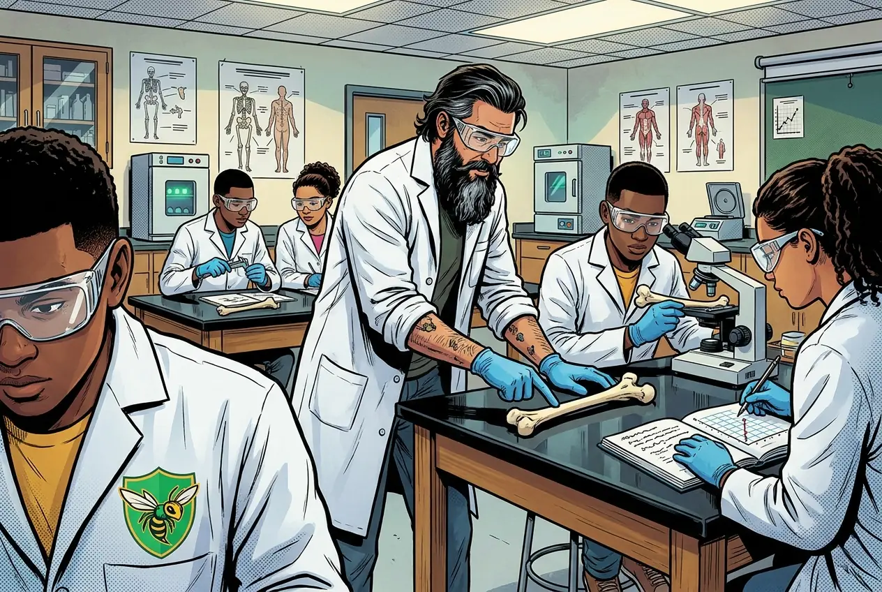
X-rays and fracture models. Greenstick, transverse, comminuted, spiral. We had to classify each break, name the nearest joint type, and describe the position using the directional terms from week one.
CLASSMATE
The bone recorded how it was broken. You are reading an event that already happened.
Fiction. There is no such student. The lessons, labs and dates are the real planned course; the student, the classmates and the conversations are invented.
🛠 Get unstuck · pick your level
Lab day: Tier 1 is the whole class at the bench. No extension today.
🔑 Today's words · 5
Tap a word in the lesson for a plain meaning and one example. Recycled into next week's Do-Now.
Do the work · 80-minute blockfirst 5 min = hook▸
💡 Big idea: Because each type of force leaves a distinct break pattern, you can read a from imaging and reconstruct how the injury happened, so classification is evidence, not labeling.
- 0-8 and lab setup; review -type reference card
- 8-20Examine X-ray set or model: classify and record
- 20-45Complete classification table with type and nearest
- 45-60Predict healing speed for each with written justification
- 60-75Group comparison: resolve disagreements using reference
- 75-80Submit classification table
- • Today is a lab day. You will look at real or simulated X-ray images and classify what you see.
- • Radiologists use the exact vocabulary you read last night. Your job is to practice that same classification process.
- • For each image you will name the type, name the nearest , and predict which would heal fastest. All three go in your .
- • Work carefully and use your references. Accuracy matters more than speed.
- • types: greenstick (incomplete, pediatric), (perpendicular break), comminuted (shattered into fragments), spiral (twisting force).
- • types near common sites: hinge (elbow, knee), ball-and-socket (hip, shoulder), pivot ( radioulnar).
- • healing speed depends on bone type, blood supply, patient age, and fracture complexity.
Unit 1.1 Bones: Bone structure/function, skeletal system, fractures, bone remodeling, repair technologies. · analysis lab
Day 3 of this lesson. Open this exact section in myPLTW (find it in Clever, Microsoft sign-in), then do the work below.
Do this: Complete the -analysis task in Lesson 1.1 Beginning with Bones on myPLTW alongside your in-class imaging work; finish online items during the classification window.
Mark the -analysis task complete after submitting your classification table.
Bone- task is done; today the task should show complete alongside your submitted .
myPLTW completion status plus your classification table.
The official PLTW activity stays inside myPLTW. If myPLTW will not open, use F1 and E1-E3 on this page to complete today's local evidence decision, then make up the official activity when access returns. Turn this in at the drop folder with your district Microsoft sign-in, or hand it to Mr. Mendoza in class. Both count as submitted. Doing the activity in myPLTW does not.
Check things off as you work, then submit. This tells Mr. Mendoza how you're doing so he can help the class. It does not replace turning in your producible through the submission route shown below.
Use the code Mr. Mendoza gave you, not your name. Saved on this device.
Unit 1.1 Bones: Bone structure/function, skeletal system, fractures, bone remodeling, repair technologies. · Fracture analysis lab
Complete the -analysis task in Lesson 1.1 Beginning with Bones on myPLTW alongside your in-class imaging work; finish online items during the classification window.
Bone- task is done; today the task should show complete alongside your submitted .
This is how Mr. Mendoza sees the class keeping pace with PLTW. Be honest, it only helps if it is accurate.
🎯 Analyze types and movement using imaging or model data.
- Review the -type reference: greenstick, , comminuted, and spiral.
- Examine the provided X-ray set or model and classify each break.
- Identify the nearest each and its type (hinge, ball-and-socket, pivot).
- Record which would likely heal fastest and why.
- Submit your classification table with notes.
Data table: classification table: each image labeled with fracture type, nearest type, and predicted healing speed with justification.
Turn this in at the drop folder with your district Microsoft sign-in, or hand it to Mr. Mendoza in class. Both count as submitted. Doing the activity in myPLTW does not. Use the checklist just below and upload by 11:29 PM for full credit. Absent with an excused absence? You get two school days for every day you were absent, so this deadline moves with you.
| Task | Who |
|---|---|
| Review the -type reference: greenstick, , comminuted, and spiral. | _______ |
| Examine the provided X-ray set or model and classify each break. | _______ |
| Identify the nearest each and its type (hinge, ball-and-socket, pivot). | _______ |
| Record which would likely heal fastest and why. | _______ |
| Submit your classification table with notes. | _______ |
Working solo? Put your own name in "Who" for every row.
- You can classify common types from an image.
- You can identify types near an injury.
- 1Do thisAnalyze fracture types and joint movement using imaging or model data.
- 2Use this resource
- 3Submit thisData table: Fracture classification table: each image labeled with fracture type, nearest joint type, and predicted healing speed with justification.
- 4Submit it here
- 1Open the drop folder.
- 2Sign in with your district Microsoft account, not a personal one.
- 3Upload the file, named Lastname_Firstname__Assignment Title.
- 4Your own upload panel says Uploaded with a green check: that is your receipt.
Turn this in at the drop folder with your district Microsoft sign-in, or hand it to Mr. Mendoza in class. Both count as submitted. Doing the activity in myPLTW does not. Human Anatomy & Physiology (Human Body Systems) › Unit 1.1 Bones: Bone structure/function, skeletal system, fractures, bone remodeling, repair technologies. › Data tableOpen the drop folder
Learn it · deck, reading, and vocabulary▸
The deck carries the prior idea forward, lets you inspect an analogy, maps the rule to biology, and ends with the same evidence decision and exit ticket used on this page.
Generated from this lesson's canonical data with a red-team citation check.
Because grafts are scarce, society must pick an allocation principle, and each principle helps one group of patients only by moving another group further back in line.
Because each type of force leaves a distinct break pattern, you can read a from imaging and reconstruct how the injury happened, so classification is evidence, not labeling.
A bridge prototype is tested against a load limit, cost limit, and user need before revision.
- Which requirement is a criterion?
- Which limit is a constraint?
- What test result should trigger a redesign?
A design improves when evidence is compared with explicit criteria and constraints.
Biomedical designs also require , ethics, and biological validation beyond a physical prototype test.
- • Bridge requirements map to design criteria.
- • Load results map to E1-E3.
- • Revision maps to the next evidence-based iteration.
Driving question: Two kids fall off the same bleachers at John Hay. One X-ray shows a bone bent and cracked on one side only; the other shows a clean break twisting up the shaft. Same fall height, different fractures, why?
What you already know: Because grafts are scarce, society must pick an allocation principle, and each principle helps one group of patients only by moving another group further back in line.
New idea: Because each type of force leaves a distinct break pattern, you can read a from imaging and reconstruct how the injury happened, so classification is evidence, not labeling.
Visual or model: F1. F1. A lesson illustration or teaching diagram for Fracture analysis lab. Use it with E1-E3; it is a model or context image, not experimental or patient data. What to notice: Trace the labeled structure, movement, or system relationship that connects form to function.
- Observe or measure the relevant feature in analysis lab.
- Organize the observation with a stable evidence ID.
- Apply this rule: A design improves when evidence is compared with explicit criteria and constraints.
- Choose the option the evidence supports and state the limit of the conclusion.
Real biomedical example: Two kids fall off the same bleachers at John Hay. One X-ray shows a bone bent and cracked on one side only; the other shows a clean break twisting up the shaft. Same fall height, different fractures, why?
What the evidence supports: E1-E3 and F1 support the daily take-home when the response meets the stated success criteria.
What it cannot prove: The package does not support claims beyond this lesson's or any real patient diagnosis.
- • : A bone-building cell that produces and lays down new bone matrix, helping bones grow, heal, and stay strong.
- • : A large bone cell that breaks down and removes old bone , helping the body remodel bone and release stored calcium.
- • : The dense, hard outer layer of bone, built from tightly packed cylindrical units, that gives the skeleton most of its strength and protection.
- • : The lightweight inner bone made of a lattice of bony struts called trabeculae, often filled with marrow and found at the ends of long bones.
- • : A break or crack in a bone, ranging from a thin hairline crack to a complete break into separate pieces.
- • : The place where two or more bones meet, often allowing movement, with and ligaments holding the bones together.
- • : A tough band of that connects bone to bone, holding joints together and keeping them stable.
Use it now: Choose one decision option. Cite E1 and E3, then explain how the rule connects the evidence to your choice.
Go further, optional: The source links below are optional enrichment. Every fact required for today's local evidence decision appears in this lesson package.
types: greenstick (incomplete, pediatric), (perpendicular break), comminuted (shattered into fragments), spiral (twisting force).
Limit: E1 supplies context or an observation; it does not by itself establish the explanation.
Because each type of force leaves a distinct break pattern, you can read a from imaging and reconstruct how the injury happened, so classification is evidence, not labeling.
Limit: E2 is a teaching statement or comparison and must be checked against the task evidence.
You can classify common types from an image.
Limit: E3 supports only the result or product criterion named here; it cannot justify a broader clinical or causal claim.
PLTW-HAP-2027-02-17 · Simulated classroom evidence scenario
Your role: anatomy and physiology consultant
Decision: Your team must decide what the evidence from analysis lab supports before submitting the labeled and result claim named on the lesson page.
- • Keep the current design.
- • Revise the feature that misses a criterion.
- • Run one more fair test before choosing.
Response: State one choice, cite at least two evidence IDs, explain the rule that connects them, and add one limitation. Submit it as the labeled and result claim.
Claim ceiling: The supplied lesson evidence can support an observation, pattern, classroom mechanism, or next-step decision about analysis lab. It cannot by itself prove causation, establish a real clinical diagnosis, or justify action outside this classroom task.
Reason for review: Your team must decide what the evidence from analysis lab supports before submitting the labeled and result claim named on the lesson page.
Context: A pattern is a record of the force that caused it, so reading breaks and the joints beside them lets you reconstruct the mechanism of injury from imaging without ever seeing the event.
- • T1: Review the -type reference: greenstick, , comminuted, and spiral.
- • T2: Examine the provided X-ray set or model and classify each break.
- • T3: Identify the nearest each and its type (hinge, ball-and-socket, pivot).
- • T4: Record which would likely heal fastest and why.
- • T5: Submit your classification table with notes.
- • E1: types: greenstick (incomplete, pediatric), (perpendicular break), comminuted (shattered into fragments), spiral (twisting force).
- • E2: Because each type of force leaves a distinct break pattern, you can read a from imaging and reconstruct how the injury happened, so classification is evidence, not labeling.
- • E3: You can classify common types from an image.
Measurements: Use only the measurements, units, graph, or counts supplied in today's task. No additional patient measurement is implied.
Figure finding: Teaching diagram for analysis lab. Trace the labeled structure, movement, or system relationship that connects form to function. This is a teaching model, not patient or experimental data.
Uncertainty: This is a composite classroom scenario. Missing history, measurements, or confirmation tests remain unknown and limit the conclusion.
Mean = sum of values / number of values. Median = middle ordered value. Range = maximum - minimum.
For 2, 4, 4, and 10: mean = 20 / 4 = 5, median = 4, and range = 10 - 2 = 8.
Mean, median, and range keep the measurement unit. Order the values before finding the median.
Calculate the requested summary for today's supplied values, then write what it reveals and what it hides.
Students often think Students think 'a break is a break,' so a just means the bone failed and the type is cosmetic detail.. The trap: type encodes the direction and nature of the force: a spiral means twisting, a greenstick means a young flexible bone bent, comminuted means high-energy shattering, so the pattern is diagnostic, not decorative, because it tells clinicians (and investigators) how the injury happened. Ignoring the type throws away the evidence.
Fracture classification table (fictional image set):
- Image 1: greenstick fracture (incomplete, bent on one side). Nearest joint: wrist (condyloid). Likely fast healing because it is incomplete and typical of younger bone.
- Image 2: transverse fracture (break straight across, perpendicular to the bone). Nearest joint: elbow (hinge). Moderate healing time.
- Image 3: comminuted fracture (bone shattered into several fragments). Nearest joint: hip (ball-and-socket). Likely slowest healing because of the many fragments.
- Image 4: spiral fracture (twisting break angling around the bone). Nearest joint: shoulder (ball-and-socket). Moderate to slow healing.
Fastest healer and why: Image 1 (greenstick), because the bone is only partly broken and is the kind of break seen in younger patients with strong blood supply, so less new bone must form.
| Image | Fracture type | Nearest joint (type) | Predicted healing |
|---|---|---|---|
| 1 | Greenstick | Wrist (condyloid) | Fastest, incomplete break |
| 2 | Transverse | Elbow (hinge) | Moderate |
| 3 | Comminuted | Hip (ball-and-socket) | Slowest, many fragments |
| 4 | Spiral | Shoulder (ball-and-socket) | Moderate to slow |
This model shows the level of evidence and organization needed to complete: A data table that classifies each fracture image by type, names the nearest joint and its type, and predicts which break heals fastest with a justification.
- Name the variables and include units.
- Enter observations without changing the raw values.
- Check labels, calculations, and patterns before interpreting the data.
Keep the structure. Replace the question, facts, measurements, and evidence. Then recheck units, vocabulary, and whether the conclusion goes beyond the evidence.
Also due today: Submit your completed data table as a photo or typed document.
- CER:
- Claim, Evidence, Reasoning: make a claim, back it with evidence, explain your reasoning.
- SOP:
- Standard Operating Procedure, the exact steps to follow (especially in a lab).
- Tracker:
- Your PLTW progress log where you record completed evidence.
- myPLTW:
- The PLTW course site where you do the online activities. Find it in Clever with your Microsoft sign-in, right next to Schoology.
Tap the speaker to hear a term. Add two of these to your notebook glossary with a definition and an example in your own words.
Pick just 2 or 3 words from today and make them yours: write what each one means in your own words, name the context clue or evidence that helped, then give one example from what you actually did in Fracture analysis lab. Try your own words first; the glossary is there if you get stuck. This is voluntary and counts as extra credit, so keep it short.
Saved on this device. Show Mr. Mendoza or add these to your notebook glossary to claim the extra credit.
Hand-picked readings, videos, and interactives for this lesson, all free and from authoritative open organizations (NIH, CDC, OpenStax, Khan Academy, PhET, HHMI, and more).
A fillable, Cornell-style notebook for Unit 1: Road to Rehabilitation. Type your notes, cues, and summaries right in the PDF, or print it and write by hand. Each lesson page has a cue column, a notes column, and a summary box, plus dated lab-record pages you can turn in.
HBS Unit 1 notebook: Road to Rehabilitation Fillable PDFCornell notes + lab recordsOpenVetted readings and references for this unit. Use them to prepare, to catch up if you were absent, or to go deeper on today's target.
Check yourself · commit, then reveal▸
Claim ceiling for this check: The supplied lesson evidence can support an observation, pattern, classroom mechanism, or next-step decision about analysis lab. It cannot by itself prove causation, establish a real clinical diagnosis, or justify action outside this classroom task.
An X-ray shows a spiral fracture of the tibia. What kind of force most likely caused it, and name one joint near that break and its type.
Write an answer and pick a confidence to unlock the key.
Fast retrieval with instant answers, not the commit-then-reveal check above. Try each from memory first: write what you remember about the earlier units, then check yourself here.
Go further and get help▸
I can name the procedure's purpose and the evidence I will record. I can identify each named hazard and the control that reduces it: If handling physical bone models, inspect for sharp edges and wear gloves if directed by the teacher. My data table is ready before materials are handled.
Finish the checklist before you handle any material.
- • If handling physical bone models, inspect for sharp edges and wear gloves if directed by the teacher.
- • Digital X-ray sets require no additional PPE but treat images with patient dignity.
- 1Before materials are handled, identify the purpose, variables or comparison, controls, measurement units, and stop-work condition.
- 2Review the fracture-type reference: greenstick, transverse, comminuted, and spiral.
- 3Examine the provided X-ray set or fracture model and classify each break.
- 4Identify the joint nearest each fracture and its type (hinge, ball-and-socket, pivot).
- 5Record which fracture would likely heal fastest and why.
- 6Submit your fracture classification table with joint notes.
- 7Record each result in the prepared table before interpreting it. Mark missing, repeated, or invalid results truthfully.
- 8Complete the named cleanup and waste route, remove PPE safely, wash hands when required, and confirm the station is ready for the next group.
| Trial or sample ID | Independent condition | Measured result with units | Observation before interpretation | Quality-control note |
|---|---|---|---|---|
Before the procedure, predict the result and cite the rule behind the prediction.
After the procedure, compare the result with the prediction and name one limitation or source of uncertainty.
What today's skills lead to. These are real health-science careers this course builds toward. Tap one to see, on the US Department of Labor's O*NET site, what the job actually involves, what it pays, and how fast it is growing.
Use the linked and X-ray resource to classify a set of provided break images by type and name the nearest for each, then submit the classification table.
MedlinePlus: FracturesThen submit your Data table. Turn this in at the drop folder with your district Microsoft sign-in, or hand it to Mr. Mendoza in class. Both count as submitted. Doing the activity in myPLTW does not.
Class still runs. Complete the online activity above (it's self-guided). Need the concept taught without a teacher? Use this authoritative explainer:
MedlinePlus: Bone Diseases and Fractures- CompleteEvery required part of the artifact is present, nothing left blank.
- AccurateThe science and the data are correct and match the evidence.
- Scientific reasoningYou explain your claim with evidence and reasoning (CER), not just an answer.
- Professional communicationClear, organized, labeled, and written the way a clinician or scientist would.
- SubmittedTurned in the right way, on the class site or handed to Mr. Mendoza in class, and confirmed. Not in Schoology: that is where the report-card grade appears later.
- Error analysis and method · counts doubleName a specific limit of the method and how it moved your result, and compare what you predicted to what happened. "Human error" does not count; say what about the procedure or instrument caused it.