
Learning Centers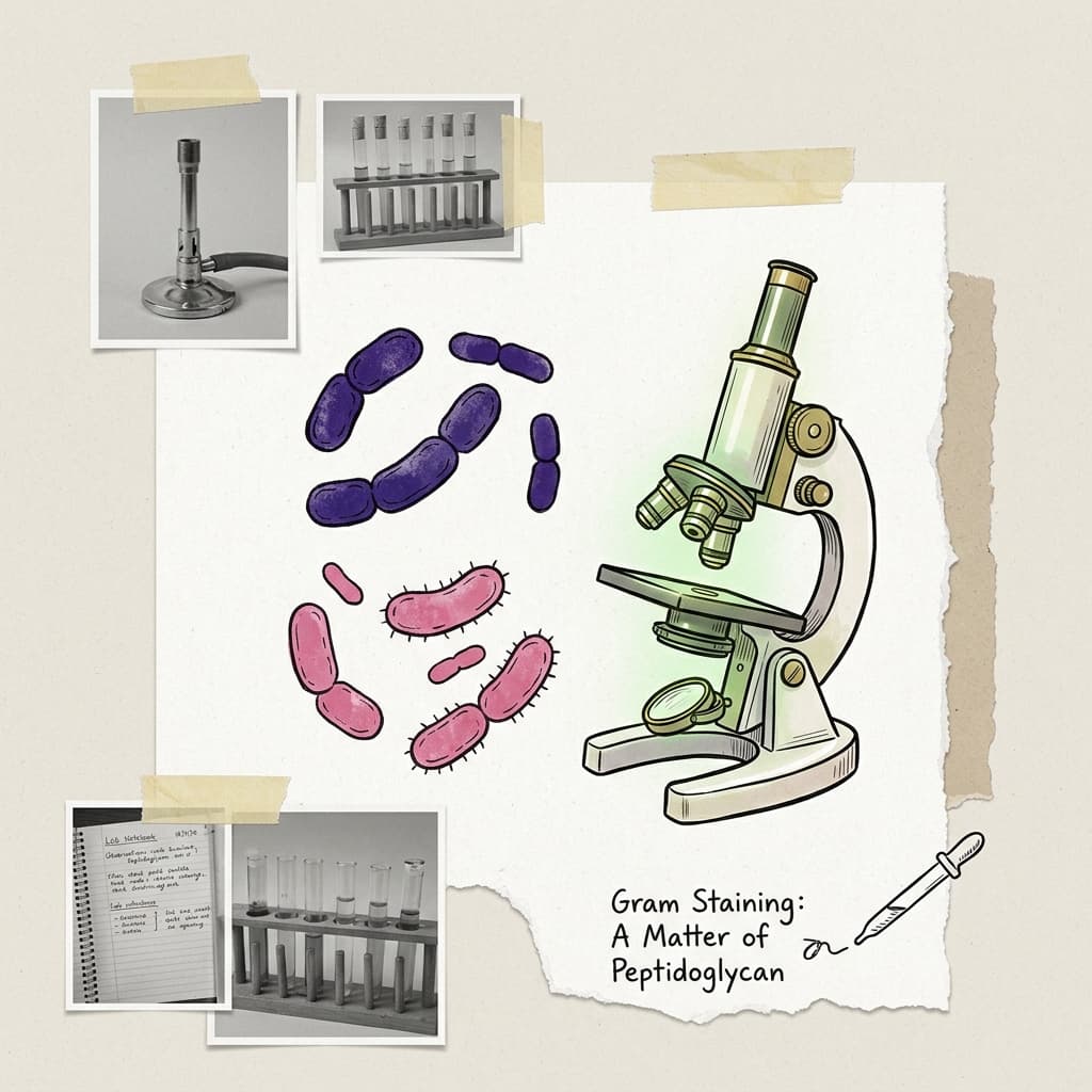
Gram Staining Bacteria
A rapid first description of an organism based on cell-wall structure.
The Two Groups
Gram-Positive
Bacteria appear purple.
Gram-Negative
Bacteria appear pink or red.
Why the Colors Differ
| Cell Type | Cell Envelope | What Happens |
|---|---|---|
| Gram-positive | Thick peptidoglycan layer | Retains the crystal violet-iodine complex during decolorization |
| Gram-negative | Thin peptidoglycan plus an outer membrane | Decolorizer disrupts the outer membrane and removes crystal violet; safranin then stains the cells |
Four Reagents and Their Jobs
- 1Crystal violet, primary stainEnters both Gram-positive and Gram-negative cells. At this point, every cell looks purple.
- 2Gram's iodine, mordantCombines with crystal violet to create a larger, less soluble dye complex inside the cells.
- 3Alcohol or acetone-alcohol, decolorizerThis is the decisive step. Gram-positive cells retain the dye complex, while Gram-negative cells lose it.
- 4Safranin, counterstainColors the now-colorless Gram-negative cells pink. Gram-positive cells remain purple because the darker crystal violet hides the safranin.