
Unit 1.3 Open Investigation: Virtual/open case investigation; synthesize scene, lab, suspect, and autopsy evidence.
Your PLTW coursework: Principles of Biomedical Science ▸ Unit 1: Medical Investigations ▸ Lesson 1.3 Open Investigation ▸ "Problem 1.3.1 Crack the Case"
What to do if absent- CER:
- Claim, Evidence, Reasoning: make a claim, back it with evidence, explain your reasoning.
- SOP:
- Standard Operating Procedure, the exact steps to follow (especially in a lab).
- Tracker:
- Your PLTW progress log where you record completed evidence.
- myPLTW:
- The PLTW course site where you do the online activities. Find it in Clever with your Microsoft sign-in, right next to Schoology.
Week overview - Open Investigation: building the evidence board and the report
Synthesize your scene, lab, suspect, and evidence into an evidence board and a that names its conclusion, reliability, and limitations.
- 1Open the Unit 1.3 project in your PLTW shell to confirm the required report sections.
- 2Gather every piece of evidence from the case and sort it on an evidence board by category.
- 3Draw connections on the board between evidence that supports the same conclusion.
- 4Write a that states your conclusion and the evidence behind it.
- 5Add a limitations section describing what your evidence cannot prove and why.
- 6Explain how an expert would defend this conclusion as reliable in testimony.
- • You can organize mixed evidence into a coherent evidence board.
- • You can write a with a clear, evidence-based conclusion.
- • You can state the limitations and reliability of your conclusion.
Daily lessons this week
Open any day for its full lesson, the work due that day, and guided notes.
Written CER (3-5 sentences) arguing what evidence threshold justifies a cause-of-death conclusion, referencing and at least one risk on either side.
Evidence planning sheet: a four-column table listing each evidence stream, the specific evidence items you have from that stream, the reliability rating, and the key limitation.
Individual notebook entry: sketch or description of your team's evidence board, the tentative cause-of-death claim, one corroborating link you found most convincing, and one gap your team could not fill.
Full investigative CER report: a clear cause-of-death claim, one piece of evidence cited from each of the four streams, a convergence reasoning paragraph, a conflict-resolution sentence, and at least two limitations.
Complete investigation packet: team evidence board photo, final CER report (claim, four-stream evidence, convergence reasoning, conflict resolution, two-plus limitations), and self-assessment form.
Quick intro to the week
- This is the unit's payoff: separate pieces of evidence become one defensible story only when you synthesize them carefully.
- Today's goal: turn your case evidence into a report whose conclusion you can defend and whose limits you can name.
- Monday's bioethics debate sharpens the work: how certain must evidence be before an expert testifies that someone is guilty?
- Your graded evidence board and are submitted on the class site.
Your PLTW coursework this week
Do this: Complete the PLTW PBS Unit 1.3 benchmark by synthesizing case evidence into an evidence board and in the online shell.
- • Synthesis combines independent pieces of evidence into a single supported conclusion.
- • Every conclusion has limitations that bound how far the evidence reaches.
- • Reliability describes how trustworthy and repeatable an evidence-based claim is.
- • Organize mixed evidence into categories on an evidence board.
- • Write a that states a conclusion and its limitations.
📋 PLTW evidence due Friday: completed Unit 1.3 evidence board and with a stated conclusion and limitations.
All PLTW activities are completed inside the PLTW course environment: this page only gives direction.
This week's PLTW tracker
Your week at a glance. Check off each deliverable as you finish it, then submit so Mr. Mendoza can see how the class is pacing.
Use the code Mr. Mendoza gave you, not your name. Saved on this device.
| Day | Date | Focus | Key deliverable |
|---|---|---|---|
| Monday | Mon, Sep 28 | Ethics of conclusions | Written CER (3-5 sentences) arguing what evidence threshold justifies a cause-of-death conclusion, referencing convergent evidence and at least one risk on either side. |
| Wednesday | Wed, Sep 30 | Synthesize all evidence | Evidence planning sheet: a four-column table listing each evidence stream, the specific evidence items you have from that stream, the reliability rating, and the key limitation. |
| Thursday | Thu, Oct 1 | Build the evidence board | Individual notebook entry: sketch or description of your team's evidence board, the tentative cause-of-death claim, one corroborating link you found most convincing, and one gap your team could not fill. |
| Friday | Fri, Oct 2 | Write the report CER | Full investigative CER report: a clear cause-of-death claim, one piece of evidence cited from each of the four streams, a convergence reasoning paragraph, a conflict-resolution sentence, and at least two limitations. |
| Friday | - | Submit investigation report | Complete investigation packet: team evidence board photo, final CER report (claim, four-stream evidence, convergence reasoning, conflict resolution, two-plus limitations), and self-assessment form. |
- M: Philosophy for Kids / John Carroll bioethical debate
- T: teacher background notes + PLTW launch task
- W: lab / data or model work
- Th: analysis / CER or design revision
- F: submit tracker + weekly evidence
Due by week's end: Evidence board and autopsy report.
5 panels from the illustrated semester.
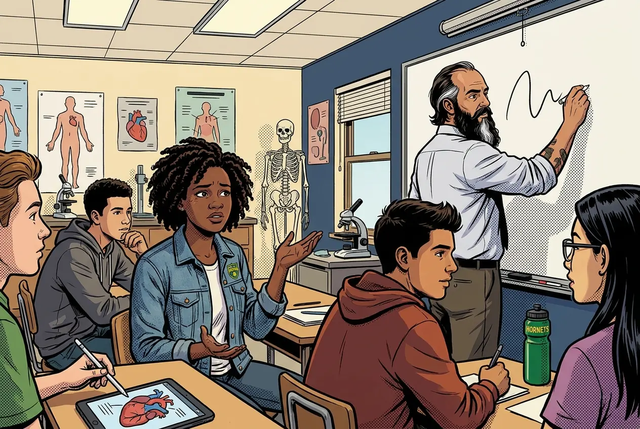
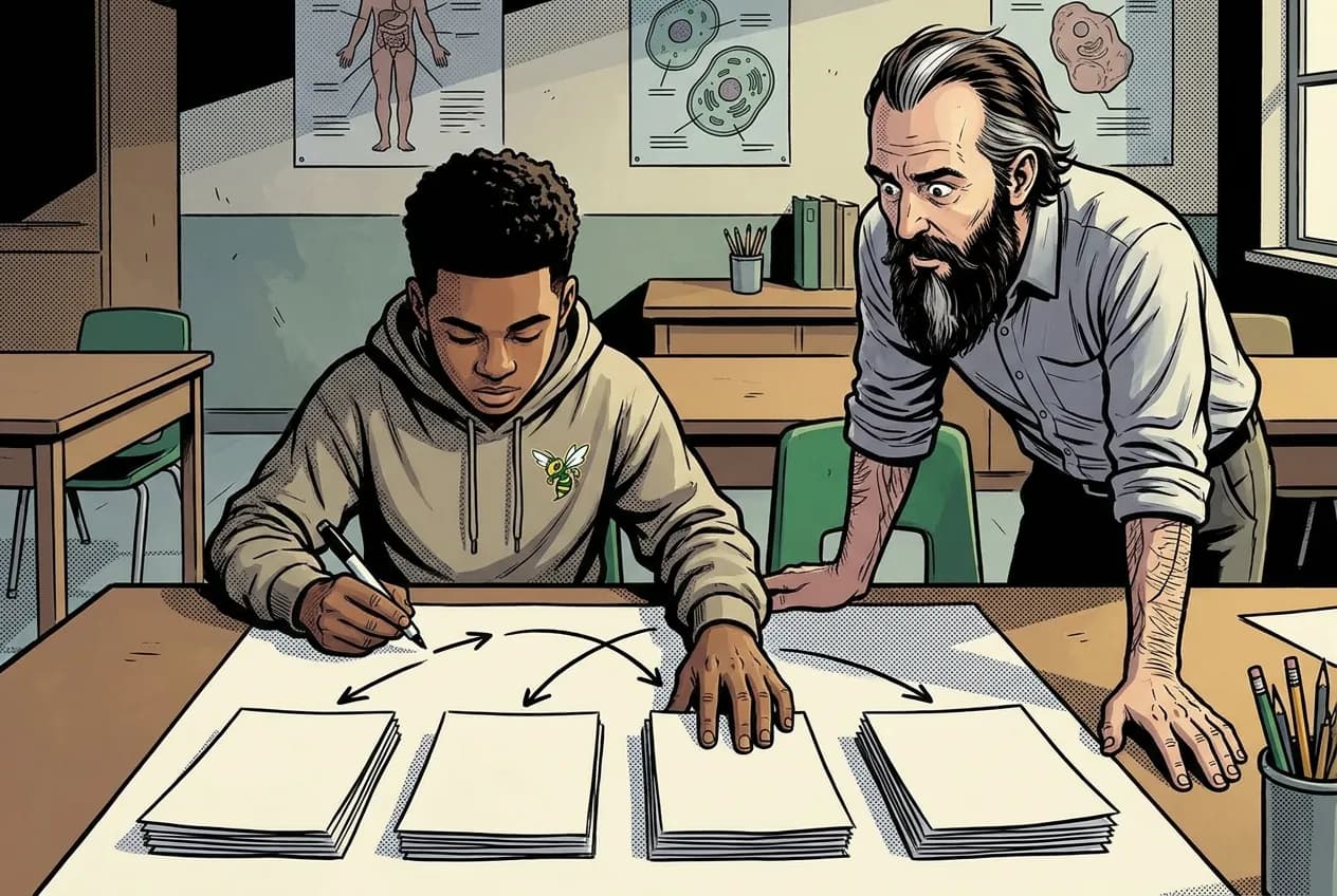
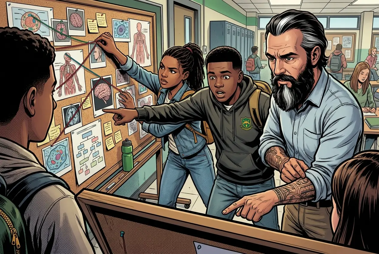
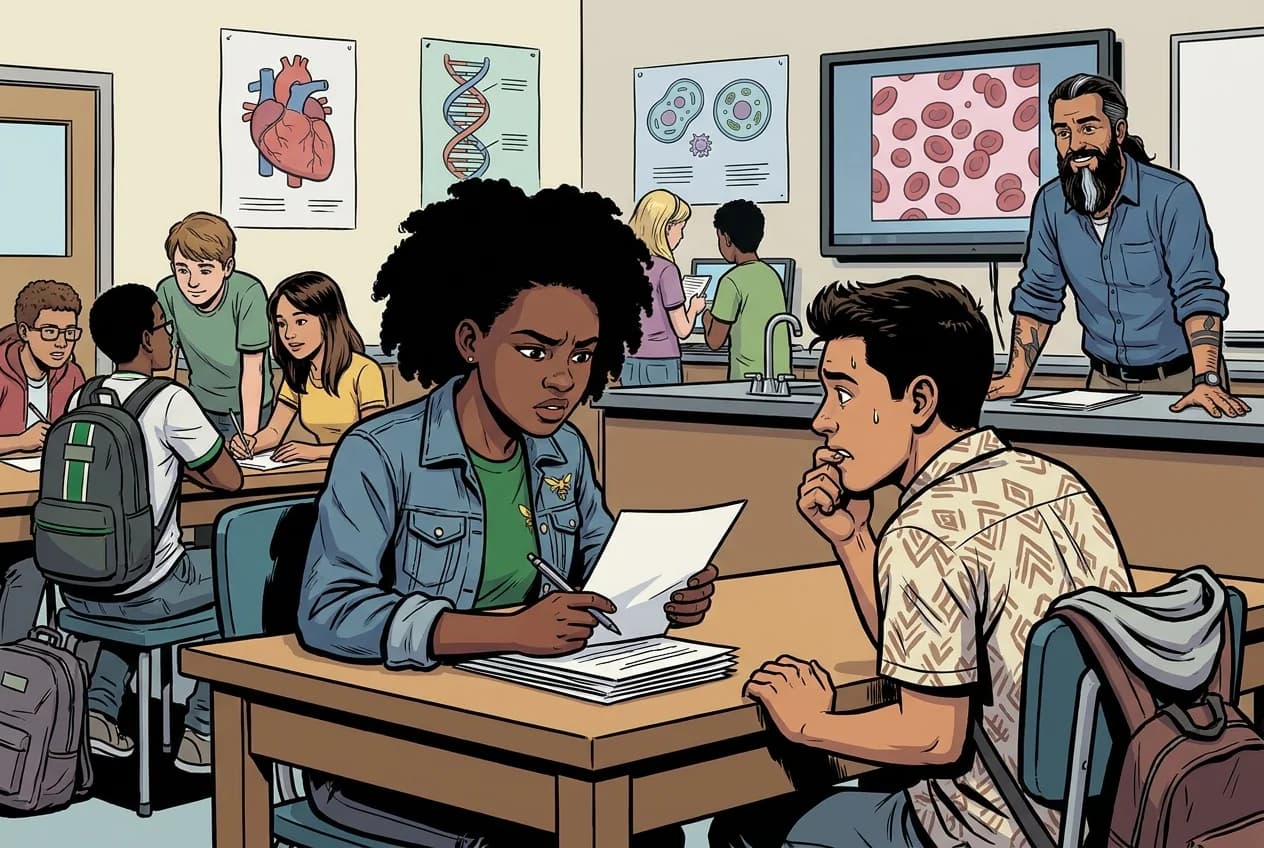
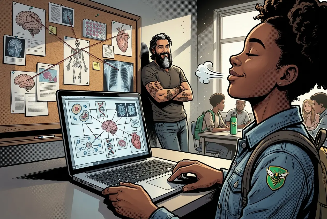
Fiction. There is no such student. The lessons, labs and dates are the real planned course; the student, the classmates and the conversations are invented.
What to do when absent
Most days, this class is your PLTW coursework: and PLTW is online and individual. So being out usually just means doing exactly what we did in class, from home.
Open Clever, then myPLTWHow to get there: open Clever and sign in with your Microsoft (district) account. Both myPLTW and Schoology are in Clever. Do the activity in myPLTW. Turn the work in on this site or hand it to Mr. Mendoza, because that is the step that counts as submitted. Schoology only shows your report-card grade later.
You can't do those from home: do this instead: Teacher-posted data/model packet, same objective. Supplemental: Khan: CER/scientific explanations; local PLTW evidence board template.
Class still runs. A substitute will post today's plan: complete the online activity above; it's built to be self-guided. Need the concept taught without a teacher? Use this authoritative explainer:
Khan Academy Biology LibraryVocabulary
Resources & readings
Hand-picked readings and interactives for this lesson, from authoritative open organizations and PLTW's own public course outline.